Science Study Guide for the TEAS
Page 3
Human Anatomy and Physiology: The Digestive, Nervous, and Muscular Systems
Anatomy and Physiology of the Digestive System
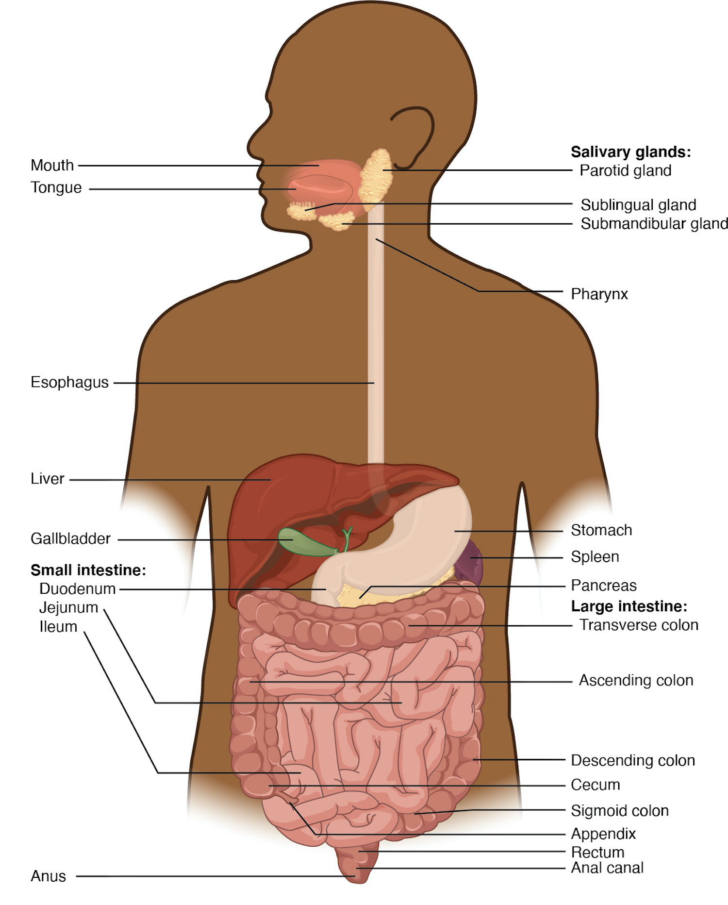
Another name for this system is the gastrointestinal system. The digestive system’s function is to provide the body with food molecules. Some of these will be broken down for energy, and some are reassembled into structures the body needs for growth and repair. It also absorbs water and other vitamins and minerals essential for body processes.
Structure

Retrieved from: https://openstax.org/books/anatomy-and-physiology/pages/23-1-overview-of-the-digestive-system
The digestive system is a large system of organs composed of the mouth, esophagus, stomach, small intestine, large intestine, pancreas, liver, gallbladder, rectum, and anus. The one-way pathway through which food travels is called the alimentary canal. The alimentary canal begins at the mouth, which breaks food apart and guides it into the esophagus. The stomach secretes digestive enzymes and churns food to break it down. It then passes through the duodenum into the small intestine, where nutrients are absorbed into the bloodstream to be delivered to cells. Finally, the large intestine absorbs water and forms waste to excrete from the anus. The salivary glands, liver, gallbladder, and pancreas are accessory organs, which excrete substances to aid in the breakdown of food.
Function
The digestive system’s function is to break down food and absorb nutrients. During digestion, the body secretes several enzymes that help break down food. These include pepsin, which acts on proteins, lipase, which acts on fat, and amylase, which acts on carbohydrates. Generally, food is moved through the digestive system via a series of muscular contractions called peristalsis.
Chemical vs. Mechanical Digestion
Food must be broken down before it can be absorbed into the bloodstream and used by cells. Mechanical digestion includes processes that break food into smaller pieces without changing it chemically. The teeth are the first structures involved in mechanical digestion, which continues when the food enters the stomach and is churned and mixed with digestive juices. Chemical digestion refers to processes that chemically change food molecules, breaking them down into smaller parts that can be used by cells. Saliva in the mouth begins this process. Stomach acid and the enzymes secreted by the liver and pancreas aid in chemical digestion.
The Role of Hormones and Secretions
The stomach produces gastric juice, a combination of hydrochloric acid and several enzymes that help break down food. The production of gastric juice is regulated by hormones and is stimulated by factors such as the smell or thought of food, as well as the presence of food itself. The pancreas secretes amylase, lipase, nuclease, and several proteolytic (protein-splitting) enzymes. The liver creates bile, which is stored in the gallbladder until needed. This substance helps enzymes break down fats. Other important functions of the liver include removing toxins and aiding in metabolic processes.
Anatomy and Physiology of the Nervous System
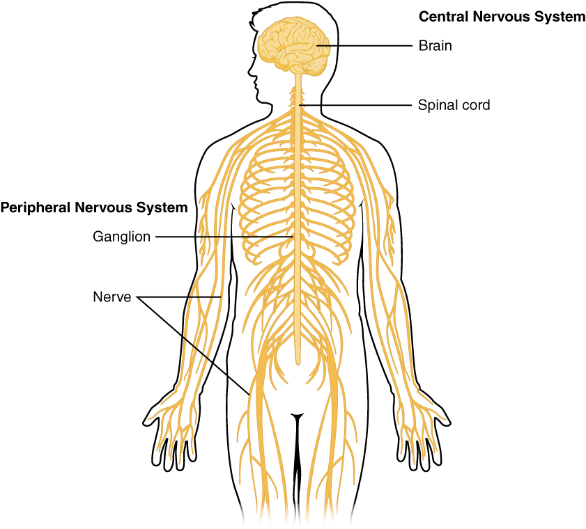
The nervous system is composed of the central nervous system, which contains the brain and spinal cord, and the peripheral nervous system, which contains the nerves and sensory organs. Some references combine the previous two systems into one, which is then known as the neuromuscular system. The brain is the control center of the nervous system and contains roughly 100 billion neurons.The nervous tissue is composed of two kinds of cells: neurons or “nerve cells,” which transmit electrochemical signals to the body, and neuroglia, which surround and help to maintain the neurons.

Retrieved from: https://openstax.org/books/anatomy-and-physiology/pages/12-1-basic-structure-and-function-of-the-nervous-system
The Central Nervous System
The central nervous system (CNS) includes the brain and spinal cord. The brain controls sensation and perception, thinking, and movement. The regions of the brain include the left and right cerebral hemispheres, the diencephalon, the cerebellum, and the brainstem. The spinal cord extends from the base of the skull through the vertebral column and ends between the first and second lumbar vertebrae at the base of the spine.
The Peripheral Nervous System
The peripheral nervous system (PNS) consists of nerves that branch from the CNS to other parts of the body. It includes cranial nerves that come from the brain and spinal nerves that come from the spinal cord. It is divided into two parts: the somatic nervous system, which controls voluntary and conscious activities, and the autonomic nervous system, which controls involuntary, subconscious activities, such as the heartbeat and digestion.
Structure of the Neuron
The structure of a neuron helps it serve its function in relaying messages throughout the body. Its structure includes a cell body, where the nucleus is found, dendrites, which are projections that receive messages from other cells, and a long axon that transmits messages to cells.
Function of the Neuron
There are three kinds of neurons: afferent neurons, which send sensory signals to the central nervous system, efferent neurons, which send signals from the central nervous system to the muscular system, and interneurons, which form the network that transmits information from afferent neurons to efferent neurons.
Anatomy and Physiology of the Muscular System
The muscular system works in conjunction with the skeletal system to allow movement. These movements include voluntary, conscious movements, such as walking or speaking, and involuntary movements, such as breathing or digestion. All muscle cells are composed of muscle fibers. Muscle cells are stimulated by the nervous system to contract as needed.
Types of Muscle Tissue
There are three types of muscle in the human body: cardiac, smooth, and skeletal.
-
Skeletal muscle is the most abundant and is, for the most part, controlled voluntarily. Tendons attach skeletal muscles to bones to create motion when the muscles contract.
-
Smooth muscle appears less striated than skeletal muscle when viewed with a microscope. It is found in the digestive tract, urinary tract, and uterus. It moves substances through these organs in a wavelike motion called peristalsis.
-
Cardiac muscle is only found in the heart.
Skeletal
Skeletal muscles, such as the deltoid and biceps brachii, are the voluntary muscles attached to bones by tendons.
Smooth
Smooth muscle is involuntary and is the stretchiest type of muscle. It is found throughout the body, in blood vessels, the bladder, the eyes, and many other areas.
Cardiac
Cardiac muscle is found in the heart and is also an involuntary muscle.
Structure
The muscle fibers that make up muscle tissues are long, thin tubes that contain contractile fibers. Muscles are made up of several bundles of muscle fibers, or fascicles. They are surrounded by connective tissue that fuses at the ends of muscles to form tendons that attach them to bones.
Function
Muscles contract in response to nerve impulses, whether voluntary or involuntary. Neurons control the contraction of cells by releasing chemicals called neurotransmitters that stimulate the fibers to contract. Motor neurons control the voluntary movements of skeletal muscles. Smooth and cardiac muscles are controlled by the autonomic nervous system. Every motion in the body is created by the work of some type of muscle.
All Study Guides for the TEAS are now available as downloadable PDFs