Multisystem Study Guide for the CCRN
Page 2
Healthcare-Associated Infections
Healthcare-associated infections, also known as healthcare-acquired infections, are infections that occur while a patient is in the hospital. Patients with invasive equipment needs are at high risk for these types of events. These infections are highly preventable and are tracked by many healthcare facilities to reduce the incidence of occurrence. Understand each of the following healthcare-associated infections, how to prevent them, and how to treat them in preparing for your exam.
Central Line-Associated Bloodstream Infections (CLABSI)
A central line-associated bloodstream infection (CLABSI) is defined as an infection that is introduced via a central venous catheter. It can be diagnosed when lab work confirms a new bloodstream infection at least two days following the insertion of a central line catheter. Central venous catheters in the intensive care unit are common and used for antibiotics, fluids, parenteral nutrition, and other intravenous medications.
Symptoms of CLABSI include hypotension, fever, chills, erythema, edema at the site, tachycardia, purulent drainage, and pain or tenderness at the catheter site. Diagnosis can be made with laboratory tests, including blood cultures and CBC. Patients will be treated with antibiotics based on the culture results. When possible, infected catheters should be removed to prevent further or future infections.
CLABSI events can significantly impact a patient and their health. They increase a patient’s hospital stay. Infections will need to be treated with antibiotics, and other supportive medications, such as vasopressors, may also need to be used. If not treated promptly or if other complications develop, patients greatly increase their mortality rate when CLABSI occurs.
Prevention of CLABSI is crucial to improving patient outcomes and reducing healthcare costs. Prevention of infection begins with good hand hygiene. This is followed by strict regulations on catheter management, including aseptic technique while accessing and maintaining the catheter, frequent assessment of the catheter, and sterile site care when needed.
Catheter-Associated Urinary Tract Infections (CAUTI)
Catheter-associated urinary tract infections (CAUTI) occur when patients acquire a urinary tract infection following placement of a catheter into the bladder. Patients with indwelling urinary catheters in place for greater than 48 hours are at risk for developing a CAUTI.
Symptoms of CAUTI include abdominal pain, fever, urinary urgency, dysuria, nausea and vomiting, lower back pain, flank pain, fatigue, and mental status changes. Diagnosis is made via urinalysis, physical assessment, and positive urine culture. Patients with positive urine cultures will need to be treated with bacteria-sensitive antibiotics. Removal of indwelling urinary catheters as soon as possible greatly reduces the risk of CAUTI.
While managing the catheter, nurses must pay careful attention to reduce the risk of infection. Prior to catheter insertion, nurses should practice hand hygiene and prepare the patient in an aseptic fashion, ensuring that all equipment maintains sterility as it is placed. Indwelling catheters should be maintained with a closed drainage system and be secured to the patient to prevent pulling or tension on the catheter. Finally, catheters should be removed as soon as medically possible to reduce the risk of infection from the indwelling device.
Ventilator-Associated Events and Pneumonia (VAE/VAP)
Patients who have to be ventilated require extra attention to prevent ventilator-associated events. Ventilator-associated events can occur when patients are intubated and mechanically ventilated for greater than 48 hours. Aspiration is one of the most common complications in patients with ventilators. This increases a patient’s risk for pneumonia due to the inability to clear secretions on their own.
Patients at highest risk for pneumonia include increased age, those intubated for an extended period of time, immobility, and those with immunocompromise. Symptoms of ventilator-associated pneumonia include fever, increased secretions, respiratory distress, hypoxemia, and cough. Diagnosis can be made with chest x-ray and respiratory secretions. This can help to determine if infiltrates are present. Treatment of ventilator-associated pneumonia (VAP) begins with the initiation of antibiotics. When an organism is identified from a sputum sample, specific antibiotics can be used to treat the organisms.
Prevention of a ventilator-assisted event (VAE) and VAP is essential to improving patient outcomes. Patients on mechanical ventilation should have their head of bed elevated to a minimum of 30 degrees. Mouth care should be performed every 2-4 hours to prevent excess secretion presence and to cleanse the mouth organisms that precipitate pneumonia. In-line suctioning and suction tubing should be changed frequently to reduce buildup of bacteria. Finally, patients should be mobilized early to prevent deconditioning and encourage quicker return of improved respiratory function.
Infectious Diseases
Infectious diseases can be the reason why a patient is in the critical care unit and/or complicate a patient’s hospitalization. Reducing patients’ exposures to multidrug-resistant organisms is a big part of improving patient outcomes and treatment. Other infectious diseases, such as influenza, pose great risk to the critically ill patient. We will review these conditions throughout this section.
Multidrug-Resistant Organisms
Multidrug-resistant organisms are becoming more prevalent in healthcare facilities. Patients who have complicated courses with these resistant bacteria have increased risk of morbidity and mortality due to the difficulty in treating the infection. Patients with suspicion of a drug-resistant organism or who have a known drug-resistant organism should be placed on strict isolation precautions to help prevent the spread of infection from patient to patient.
MRSA
MRSA, or methicillin-resistant staphylococcus aureus, is a type of bacteria that causes 10-50% of all staph infections and is resistant to many antibiotics. This makes the infection difficult to treat. Patients at increased risk for MRSA include those with frequent hospitalizations, infection development after recent antibiotic therapy, and immunosuppression.
Many community members are carriers of MRSA due to various exposures. While this does not mean they are actively infected, they do have a higher risk of acquiring and spreading MRSA bacteria to places of injury where it can become opportunistic and cause a serious infection. MRSA infection can present as a skin, urinary tract, respiratory, bloodstream, or wound infection.
Symptoms of MRSA are similar to all other infectious diseases. Patients may experience fever, pain, erythema (at the site), swelling, chills, headache, shortness of breath, fatigue, cough, improper or delayed healing, and open wound drainage. Diagnosis can be made with a culture swab of the suspected area of infection.
MRSA is treated with antibiotics. After diagnosis is made, usually MRSA antibiotic sensitivities can be determined from the culture swab. Using antibiotics that are proven to work against MRSA are critical to treating the infection. If MRSA has infected an open wound, surgical irrigation and debridement may also be required.
VRE
Vancomycin-resistant enterococcus (VRE) is another common multidrug-resistant bacteria. It is caused by Enterococcus bacteria and can be found in infections involving the urinary tract, bloodstream, endocardium, and meninges. Patients at highest risk for acquiring VRE include those with prolonged hospital stays, those on long-term antibiotics following surgical operation, and those who are immunosuppressed.
As with the infectious symptoms described above in MRSA, VRE symptoms include chills, fever, and fatigue. Patients with a urinary tract infection may have additional symptoms of back pain, urinary frequency, urgency, and/or dysuria. Wound site infections may additionally present with drainage, erythema, and edema. Diagnosis of VRE is performed via culture swab. Treatment with antibiotics is based on the culture swab to ensure appropriate antibiotic therapy is initiated.
CRE
Carbapenem-resistant enterobacteriaceae, CRE, is a less common antibiotic-resistant organism. The Klebsiella pneumoniae organism is the most common of these types of infection. Patients at increased risk include those who are mechanically ventilated, have indwelling equipment such as a urinary catheter, IV access, and those with prolonged antibiotic courses. Infections may be present in the urinary tract system or respiratory system. They may also be present in bloodstream infections or wounds.
Diagnosis of CRE can be achieved via culture swab. Upon return of the results, a panel of antibiotics will be identified to treat the infection. Antibiotic choice, then, is reserved to the appropriate therapy as determined by the lab. This helps to reduce creating further antibiotic-resistant bacteria.
Influenza
Influenza is a virus that occurs across the world. Pandemic influenza can develop when new strains of influenza develop or strains that are non-human morph into viruses that infect humans. This type of influenza spreads rapidly because humans have little to no immunity against these new strains. Symptoms of pandemic influenza are often severe and cause many to be hospitalized for treatment. Rarely do pandemic influenza outbreaks occur.
Epidemic influenza outbreaks are more common. Deaths by influenza are tracked every year. When the number of deaths exceeds the CDC’s expected percentage of deaths, it is defined as an epidemic. Epidemics can also be defined when more people are affected by the virus in a specific area or population than expected.
Symptoms of influenza include fever, chills, myalgia, headache, fatigue, and upper respiratory symptoms. Some patients may also experience nausea and vomiting; however, this is not a hallmark of the virus. When it is suspected that a patient has influenza, a rapid detection swab may be performed. Diagnosis may also be made by physical assessment.
Since influenza is a virus, treatment is often supportive. Providing adequate hydration and rest are essential to recovery. If symptoms are identified early, antiviral medication may be prescribed to lessen symptoms. Patients with severe upper respiratory symptoms may develop pneumonia or other secondary infection that requires them to be hospitalized for additional management. Influenza vaccinations may be obtained annually and may prevent or lessen symptoms in patients exposed to the virus.
Necrotizing Fasciitis
Necrotizing fasciitis is a serious and sometimes deadly infection that develops within the fascia tissues. It spreads rapidly and can cause cell death, leading to destruction of the soft tissues and nerves. Infection is introduced via an open wound. Even with prompt treatment, the rapid spread of infection and cell death can lead to amputation, sepsis, or death. Early identification of this condition is critical to reducing potential harm.
Symptoms of necrotizing fasciitis include erythema, pain, and edema at the affected site. Systemic symptoms of nausea, fatigue, vomiting, fever, and chills may also occur. Diagnosis can be made on physical exam and history. Deep skin biopsies can be utilized to identify the offending organism. The most common organisms to cause this condition are Group A Streptococcus, Klebsiella, Clostridium, Escherichia coli, Staphylococcus aureus, and Aeromonas hydrophila. Many patients will also undergo a CT scan or MRI to determine the extent of the infection and affected tissues.
Patients diagnosed with necrotizing fasciitis will be started on antibiotics geared toward treatment of identified organisms. In many cases, surgical intervention is necessary and patients will undergo a fasciotomy and debridement. This helps to relieve the pressure of the infection on the tissue as well as clean up the dead cells. Several debridements may be necessary and recovery takes time. In some cases, patients are placed in hyperbaric oxygen chambers to enhance tissue healing and growth.
Maternal and Fetal Complications
Pregnant women can develop critical complications during and following pregnancy that can affect both the mother and the baby. Women of childbearing age should be screened for pregnancy upon admission to the emergency department or critical care unit, as many medications and therapies may need to be altered with pregnancy. For women who are already pregnant, the following conditions may occur.
Eclampsia
Eclampsia is a rare and potentially fatal complication of pregnancy. Women with elevated blood pressures during pregnancy may be diagnosed with preeclampsia. Eclampsia is a complication of this condition. When the blood pressure is uncontrolled, it can increase dramatically and cause seizures.
Eclampsia most commonly occurs in the late third trimester to 48 hours postpartum. Symptoms of eclampsia include facial and hand swelling, elevated blood pressure, weight gain, headache, nausea and vomiting, blurred vision, decreased urination, proteinuria, and right upper abdomen pain. Some mothers have no symptoms, yet develop agitation, seizures, and loss of consciousness associated with eclampsia.
Risk factors for eclampsia include first pregnancy, multiple pregnancy, age less than 20 and greater than 35 years, history of hypertension, diabetes, and kidney disease. Fetuses may measure small as eclampsia hypertension reduces the placenta blood supply, decreasing the amount of oxygen delivered to the fetus. Mothers may be induced early due to placental insufficiency. In addition to seizures, inadequate management of eclampsia can lead to placental abruption. This is a medical emergency and requires an emergent c-section to deliver the baby.
Testing of women with eclampsia may include pulmonary capillary wedge pressure (PCWP) monitoring (generally low to elevated) and lab work including CBC, creatinine, and electrolyte monitoring,
Treatment for women with eclampsia focuses on safe delivery of the baby and placenta. If the fetus is not mature enough for delivery, mothers may be prescribed seizure medications, blood pressure medications, and steroids to help develop the baby’s lungs in preparation for delivery.
HELLP Syndrome
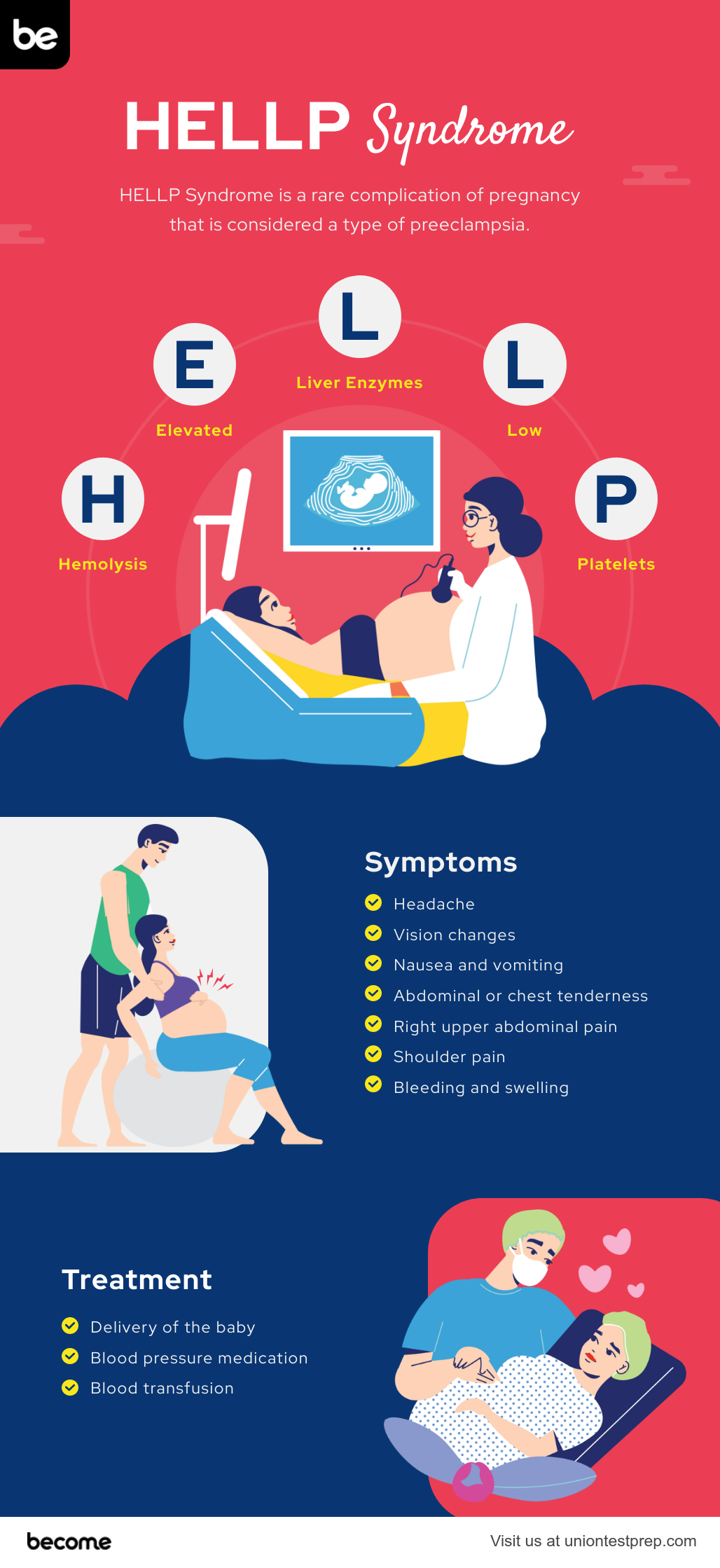
HELLP syndrome is another potential complication of preeclampsia. HELLP stands for hemolysis (H), elevated liver enzymes (EL), and low platelet count (LP). This critical condition carries a mortality rate up to 30%.
Symptoms of HELLP syndrome include headache, vision changes, nausea and vomiting, abdominal or chest tenderness, right upper abdominal pain, shoulder pain, bleeding and swelling. Most women will also experience elevated blood pressures and proteinuria. However, some may present without hypertension and proteinuria which can delay recognition of the condition. Risk of death increases significantly with liver rupture or stroke.
The Mississippi classification is used to determine the severity of HELLP. Women may fall within any of the three following classes:
- Class 1— severe thrombocytopenia with platelets less than 50,000/\(mm^3\)
- Class 2— moderate thrombocytopenia with platelets between 50,000-100,000/\(mm^3\)
- Class 3— mild thrombocytopenia (100,000-150,000/\(mm^3\)) with elevated AST greater than 40 IU/L
Treatment of HELLP focuses on delivery of the baby. Most symptoms resolve by 48-72 hours post delivery. Some women may require blood transfusions and corticosteroids to improve the lung maturity of the baby. Other medications that may be considered include magnesium sulfate to prevent seizures and antihypertensive medications. Early recognition of symptoms leads to improved outcomes for both the mother and baby. Women should be educated that risk of recurrence in subsequent pregnancies is elevated.

Postpartum Hemorrhage
Postpartum hemorrhage is one of the most common complications of delivery. It is defined as blood loss of greater than 1000 mL with symptomatic hypovolemia within 24 hours following delivery. Secondary postpartum hemorrhage may also occur greater than 24 hours following delivery. Blood loss greater than 500 mL in vaginal delivery should be investigated as this is considered abnormal.
Uterine atony, perineal or vaginal lacerations, placenta retention, uterine inversion, and coagulation disorders are all possible causes of postpartum hemorrhage. Of these, uterine atony is the most common cause. Infection and retained products of conception can also cause secondary hemorrhage. Disseminated intravascular coagulation (DIC) may present if widespread bleeding occurs from all puncture sites. Excessive blood loss can lead to Sheehan syndrome (postpartum hypopituitarism). Other complications of severe blood loss include transfusion-related acute lung injury, infections, and hemolytic transfusion reactions.
Following delivery, nurses should monitor closely for excessive bleeding from the vagina, tachycardia, tachypnea, and faint feeling with standing or repositioning. If blood loss is significant and progressive, patients may develop clamminess, hypotension, and hypovolemic shock. Regular uterine exams and bimanual massage should be performed following delivery to assess for uterine bogginess associated with uterine atony.
Common lab work associated with this condition include a type and screen, complete blood count, coagulation studies, and fibrinogen levels. Blood transfusions may be indicated if the patient’s hematocrit and hemoglobin are significantly decreased. Nurses should ensure that mothers have adequate IV access to use in the event of fluid or blood replacement.
Medications that may be used in the event of postpartum hemorrhage include oxytocin, methylergonovine, carboprost, and misoprostol. These medications are used to help induce or sustain uterine contraction. Some mothers may require uterine artery embolism if bleeding is persistent. Exploratory laparotomy is a late option for postpartum hemorrhage if more conservative measures have been ineffective.
Amniotic Embolism
Amniotic embolism is a rare, deadly complication following delivery. It occurs when amniotic fluid or fetal material enters the mother’s bloodstream. It most commonly occurs during delivery or immediately following when the placenta is disrupted or if trauma occurs.
Symptoms develop rapidly. Symptoms of amniotic embolism include sudden shortness of breath, sudden hypotension, pulmonary edema, cardiovascular collapse, DIC, acute change in mental status, impending sense of doom, tachycardia, fetal distress, seizures and loss of consciousness. When amniotic fluid enters the woman’s bloodstream, it can activate an immune response that induces abnormal clotting in the lungs and blood vessels.
Women at increased risk for amniotic embolism include those with advanced maternal age, preeclampsia, medically induced labor, c-section, placenta problems, and polyhydramnios. Amniotic embolism can result in prolonged hospital stays, brain injury, maternal and fetal death. Treatment for this condition involves oxygen administration, central and arterial line placement for cardiovascular monitoring, and cardiopulmonary resuscitation. Most women with amniotic embolism require critical care management.
All Study Guides for the CCRN are now available as downloadable PDFs