Differences Between Invasive Hemodynamic Monitoring Devices
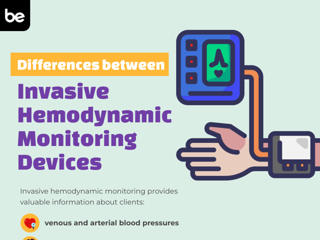
Invasive hemodynamic monitoring provides valuable information about patients’ venous and arterial blood pressures, pulmonary pressures, and cardiac output to help identify abnormal physiology that may cause organ failure and death. Critical care nurses must know differences and management strategies for each of the following devices.
Central Venous Catheter
Central venous catheters can be utilized to measure a patient’s central venous pressure (CVP). The CVP signifies the pressure within the intrathoracic vena cava. This can be used to estimate the patient’s right atrial pressure and cardiac preload. Elevated measurements (>12 mmHg) may indicate fluid overload due to increasing right atrial pressures and increased preload. Lowered measurements (<5 mmHg) may indicate hypovolemia. CVP measurements may be altered based on venous tone, intrathoracic pressure, and compliance of the heart.
Intra-Arterial Catheter
Intra-arterial catheters provide invasive, continuous blood pressure monitoring. They can be used to draw arterial blood gasses and other labs. Arterial catheters are typically inserted in the wrist or groin. These catheters are commonly referred to as A-lines.
Pulmonary Artery Catheter (Swan-Ganz Catheter)
The pulmonary artery catheter (PAC), also known as the Swan-Ganz Catheter, is utilized to monitor for fluid overload or circulatory concerns. It can be used to assess for or diagnose cardiac tamponade, heart disease, pulmonary hypertension, and cardiomyopathy.
The PAC is inserted into an arm or groin vein and advanced through the circulatory system until it enters the heart. The catheter is then floated from the right atrium to the right ventricle and into the pulmonary artery while waveforms and pressures are recorded. Once reaching the pulmonary artery, the catheter balloon is briefly inflated to occlude the artery to record a pulmonary artery wedge pressure. After the tracings are completed, the balloon is deflated and the catheter secured to the patient for reuse as needed.
Nurses using PAC devices should monitor patients closely throughout the procedure. Patients should have continuous ECG monitoring to watch for cardiac arrhythmia. Complications of the procedure may include bruising, bleeding, and pneumothorax.
Intra-Aortic Balloon Pump
The intra-aortic balloon pump (IABP) can be used to support blood circulation in patients with heart failure, cardiogenic/septic shock, and unstable angina. It can also be utilized to monitor blood pressure.
The IABP is a catheter with an inflatable balloon at the tip. It is inserted via the femoral artery and threaded into the descending thoracic aorta. By inflating the balloon during diastole and deflating during systole, the device increases circulation to the coronary artery, reducing afterload. Accurate positioning of the device is critical to help prevent side effects such as dizziness and decreased radial pulse (too high) or flank pain and decreased urine output related to renal artery occlusion (too low).
Nursing responsibilities for managing this device include:
- Hourly site inspections after the initial insertion monitoring period
- Prevent the patient from bending their knees >45 degrees
- Monitor for stroke
- Peripheral perfusion assessments
Nurses must also be prepared for device malfunctioning or damage to the balloon. Any breaking or leaking of the catheter is a medical emergency. The patient should be immediately placed into Trendelenburg and the physician notified for further management.
PiCCO Monitor
The PiCCO monitor is utilized for monitoring fluid status and pulmonary edema. The data garnered can indicate central venous pressure, cardiac preload, and overall cardiac function. It is typically used for patients with acute respiratory distress syndrome (ARDS), trauma, or shock.
PiCCO is short for Pulse index Contour Continuous Cardiac Output. Patients in need of PiCCO monitoring require intra-arterial and central venous catheterization. The machine uses two techniques to measure hemodynamic status:
- Transpulmonary thermodilution
- Pulse contour analysis
To collect data using the PiCCO, the nurse will utilize the transducer’s equipment to quickly inject cold saline into the patient’s central venous catheter which then circulates until the temperature-changed blood is then picked up on the temperature-sensitive transducer in the arterial line. The curves and data determined by this process produce the intrathoracic blood volume (ITBV), extravascular lung water (EVLW), and extravascular lung water index (EVLWI) readings.
Critical care nurses should be familiar with devices that provide hemodynamic monitoring. These devices provide essential data to help treat and prevent complications in severely ill patients. For more in-depth information on hemodynamic monitoring and other critical care concepts, or to test you knowledge, check out our CCRN practice tests, study guides, and flashcards.
Keep Reading

CCRN Blog
Is CCRN Certification Worth It?
The CCRN (Critical Care Registered Nurse) Certification stands as a tes…

CCRN Blog
Is the CCRN Exam Hard?
In the US, over 700,000 people work in critical care units (CCUs). With…

CCRN Blog
Traumatic Brain Injury and Intracranial Hemorrhage
Traumatic brain injury and intracranial hemorrhage are two common diagn…