The Heart: Five Areas for Listening
Basics of Auscultation
Auscultation of a heart begins with two critical items: a stethoscope and a patient. Knowledge about both these elements is key to assessing the health of a heart. Classic stethoscopes have two sides of the chestpiece—the diaphragm and the bell. The larger, flatter side is the diaphragm and is used for listening to higher-pitched sounds. The bell is the smaller, concave side that allows for auscultation of lower-pitched sounds like some heart murmurs. When performing a cardiac exam, auscultation should be performed with the diaphragm and then repeated with the bell.
Heart Valves
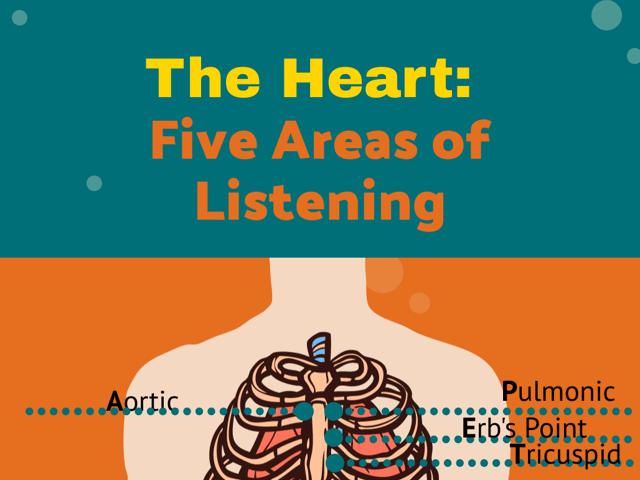
The locations of auscultation center around the heart valves. The aortic, pulmonic, tricuspid, and mitral valves are four of the five points of auscultation. The fifth is Erb’s point, located left of the sternal border in the third intercostal space. The aortic point is located right of the sternal border in the second intercostal space. The pulmonic point is to the left of the sternal border in the second intercostal space. The sound that emits from the aortic and pulmonic points is the S2 “dub” of the typical “lub-dub” heartbeat. The S1 and S2 sounds are present in normal heartbeat patterns.
The tricuspid point is found left of the sternal border in the fourth intercostal space, and the mitral point is located midclavicular on the left side of the chest in the fifth intercostal space. Both the tricuspid and the mitral points are where the S1 “lub” can be heard. The base of the heart is where the aortic and pulmonic S2 sound will be loudest. The apex is where the tricuspid and mitral S1 sound is loudest upon auscultation. The apex region will also be where S3 and S4 sounds(extra heart sounds not usually noted in normal assessments) and mitral stenosis murmurs may be auscultated, if present.
Mnemonic device
A mnemonic that aids in recalling the points of auscultation is APE To Man:
\[\begin{array}{|l|l|} \hline \mathbf{Auscultation \;Point} & \mathbf{Location} \quad \quad \quad \quad \quad & \mathbf{Sound} \quad \quad \quad \quad \quad \\ \hline \mathbf{A}\text{ortic} & \text{Right of sternal border} & \text{S2 “dub”} \\ \text{}& \text{in second intercostal } & \text{} \\ \text{}& \text{space (Base)} & \text{} \\ \hline \mathbf{P}\text{ulmonic} & \text{Left of sternal border} & \text{S2 “dub”} \\ \text{}& \text{in second intercostal} & \text{} \\ \text{}& \text{space (Base)} & \text{} \\ \hline \mathbf{E}\text{rb’s Point} & \text{Left of sternal border in} & \text{S1 and S2 (line of} \\ \text{}& \text{third intercostal space} & \text{separation between} \\ \text{}& \text{} & \text{base and apex)} \\ \hline \mathbf{T}\text{ricuspid} & \text{Left of sternal border} & \text{S1 “lub”} \\ \text{}& \text{in fourth intercostal} & \text{} \\ \text{}& \text{space (Apex)} & \text{} \\ \hline \mathbf{M}\text{itral} & \text{Midclavicular on left} & \text{S1 “lub”} \\ \text{}& \text{side of chest in fifth} & \text{} \\ \text{}& \text{intercostal space (Apex)} & \text{} \\ \hline \end{array}\]Patient positioning during assessment facilitates the auscultation of various valve anomalies. Initially, a complete auscultation assessment should be performed as the patient is in supine or sitting position. Then, patients should be positioned laterally onto their left side so the provider can listen with the bell of the stethoscope for any S3, S4 (extra heart sounds) and/or mitral stenosis murmurs in the apex region. Aortic and pulmonic murmurs are more easily identified with the diaphragm of the stethoscope when patients are in a sitting position, leaned forward, and asked to exhale.
It is important to perform a comprehensive assessment of the heart, listening to all five points and keeping in mind which side of the chestpiece should be utilized during listening, as well as the patient’s position during auscultation.

Keep Reading

National Council Licensure Examination-Registered Nurse Blog
What to Expect in Nursing School Clinicals
The clinical experience is a rite of passage for all nursing students, …

National Council Licensure Examination-Registered Nurse Blog
What is the NCLEX Next Generation (NGN) Exam?
If you’re interested in becoming a registered nurse, you likely know th…

National Council Licensure Examination-Registered Nurse Blog
IDEA for Drugs to Treat Bradycardia/Hypotension
For hospitalized patients, bradycardia (slow heart rate) and hypotensio…